Unlocking the Secrets of Radiology: Exploring the Power of Medical Imaging
Introduction
Radiology plays a pivotal role in modern medicine, enabling healthcare professionals to visualize and diagnose various medical conditions. Through the power of medical imaging, doctors can unlock a wealth of information hidden within the human body. In this article, we will delve into the fascinating world of radiology, uncovering its secrets and exploring the immense potential it holds. From X-rays to MRI scans, we will examine different imaging techniques and their applications. So, fasten your seatbelts as we embark on a captivating journey through the realm of medical imaging.
The Evolution of Radiology: From X-Rays to Cutting-Edge Technology
Radiology has come a long way since the discovery of X-rays by Wilhelm Conrad Roentgen in 1895. This groundbreaking discovery revolutionized the field of medicine, allowing doctors to visualize the internal structures of the body without invasive procedures. X-rays quickly became an invaluable tool in diagnosing fractures, identifying tumors, and detecting various diseases.
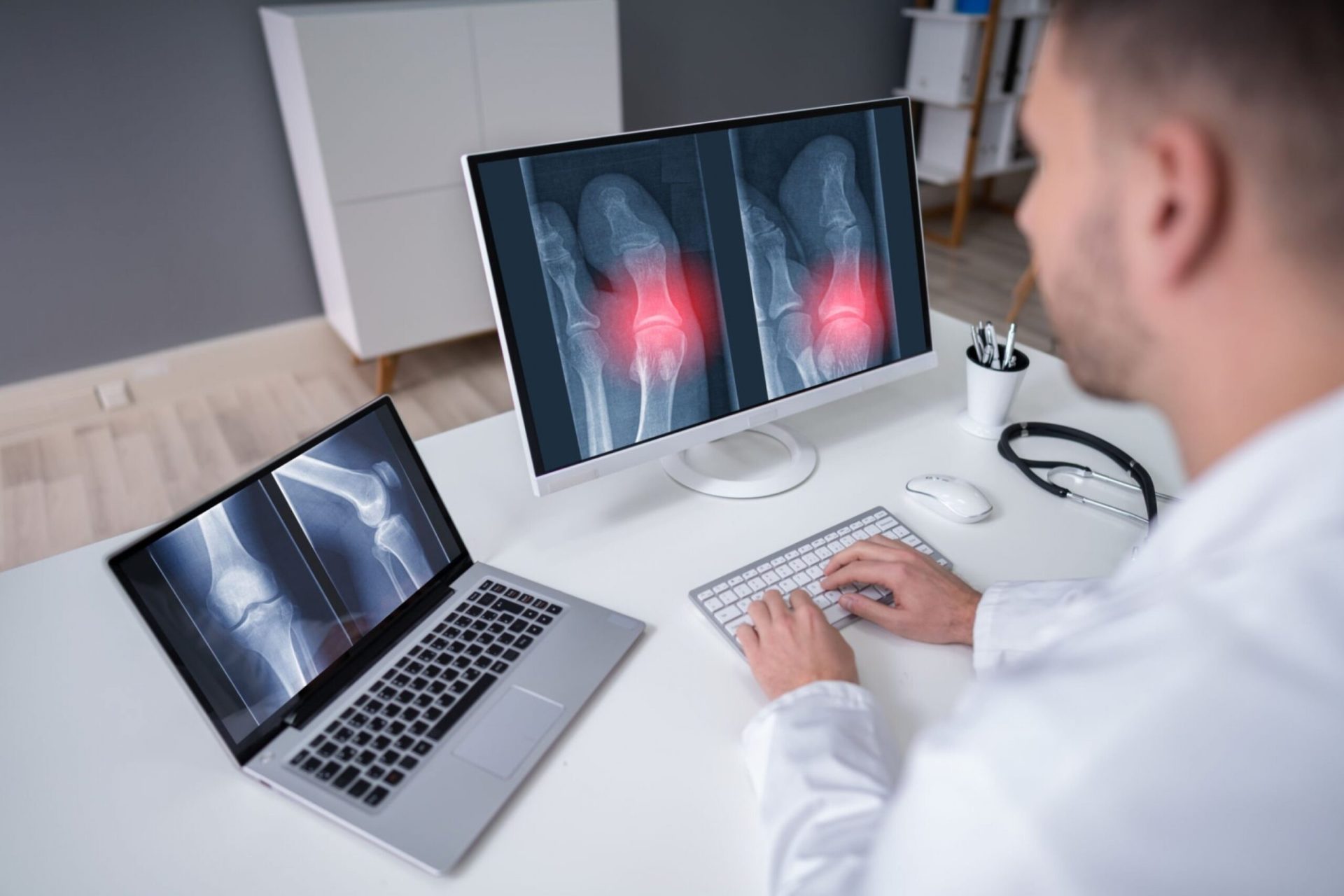
X-Rays: Peering into the Invisible
X-rays are a form of electromagnetic radiation that can pass through soft tissues but are absorbed by denser materials like bones. This property enables radiologists to capture detailed images of the skeletal system, aiding in the diagnosis and treatment of bone fractures, joint abnormalities, and other conditions.
Did you know? X-rays can also be used for dental imaging, detecting tooth decay, jaw problems, and evaluating the success of dental procedures.
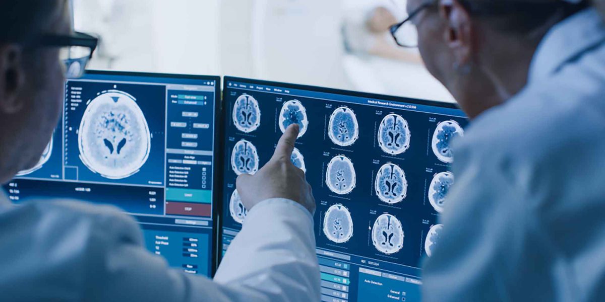
Computed Tomography (CT): Unveiling Three-Dimensional Images
As technology advanced, so did the capabilities of radiology. Computed Tomography, commonly known as CT, emerged as a powerful imaging technique that provides cross-sectional images of the body. By combining multiple X-ray images taken from different angles, a computer reconstructs detailed 3D images of internal organs, blood vessels, and other structures.
CT scans have become invaluable in diagnosing conditions such as lung cancer, cardiovascular disease, and abdominal disorders. They allow doctors to visualize intricate anatomical details and identify abnormalities with great precision.
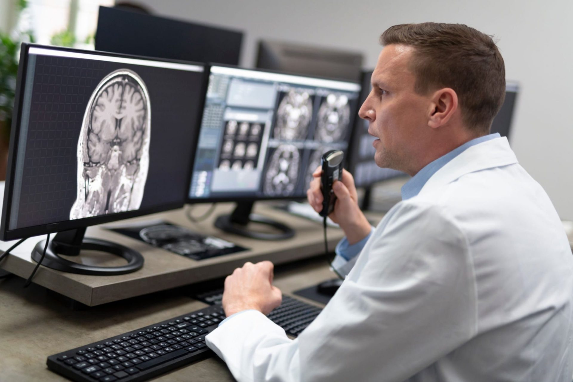
Magnetic Resonance Imaging (MRI): Peeking Inside with Magnetism
Magnetic Resonance Imaging, or MRI, harnesses the power of strong magnetic fields and radio waves to generate detailed images of the body’s organs and tissues. Unlike X-rays and CT scans, MRI does not use ionizing radiation, making it a safer option for certain patients, such as pregnant women and children.
MRI excels in capturing soft tissue details, making it particularly useful for diagnosing conditions affecting the brain, spinal cord, joints, and abdomen. It can detect tumors, lesions, and abnormalities that might not be easily visible with other imaging techniques.
Ultrasound: Harnessing Sound Waves for Visualization
Ultrasound imaging, also known as sonography, utilizes high-frequency sound waves to create real-time images of the body’s internal structures. By transmitting and receiving sound waves through a handheld device called a transducer, doctors can visualize organs, blood vessels, and developing fetuses.
Ultrasound imaging is widely used in obstetrics and gynecology to monitor pregnancies, check fetal development, and identify potential complications. It is also employed in examining the heart, liver, kidneys, and other organs, providing valuable diagnostic information.
Advancements in Radiology: Pushing the Boundaries of Medical Imaging
The field of radiology is continuously evolving, driven by technological advancements and innovative approaches. Let’s explore some of the latest developments that are revolutionizing medical imaging and unlocking new possibilities for diagnosis and treatment.
Positron Emission Tomography (PET): Illuminating Metabolic Activity
Positron Emission Tomography, or PET, combines molecular imaging with functional information, enabling doctors to observe metabolic
Common FAQs
What are the risks associated with medical imaging procedures?
Medical imaging procedures, such as X-rays, CT scans, and MRI scans, are generally considered safe. However, they do involve exposure to radiation in the case of X-rays and CT scans. The risk of radiation exposure is typically minimal and outweighed by the benefits of accurate diagnosis. Pregnant women are advised to inform their healthcare providers before undergoing any imaging procedures to ensure appropriate precautions are taken. MRI scans do not involve radiation and are considered safe for most individuals.
How do I prepare for a medical imaging procedure?
The preparation for a medical imaging procedure depends on the specific test being performed. Your healthcare provider will provide you with detailed instructions tailored to your situation. In some cases, you may need to fast for a few hours before the procedure or avoid certain medications. It is important to follow these instructions closely to ensure accurate results.
How long does a typical imaging procedure take?
The duration of an imaging procedure varies depending on the type of test and the body part being examined. X-rays and ultrasound tests are usually completed within a few minutes, while CT scans and MRI scans may take anywhere from 15 minutes to an hour or longer, depending on the complexity of the study.
Are medical imaging procedures painful?
Most medical imaging procedures are painless. However, some individuals may experience minor discomfort or claustrophobia during certain tests such as MRI scans. If you have concerns about discomfort or claustrophobia, discuss them with your healthcare provider beforehand. They can provide strategies to help you remain comfortable during the procedure.
Can medical imaging procedures be performed on children?
Yes, medical imaging procedures can be performed on children. However, special considerations are taken to ensure their safety and comfort. Radiation doses for pediatric patients are carefully adjusted to minimize exposure. Sedation or anesthesia may be used for younger children or those who have difficulty remaining still during the procedure.
Are there any alternatives to radiation-based imaging techniques?
In some cases, alternative imaging techniques can be used to avoid or minimize radiation exposure. For example, ultrasound and MRI scans are radiation-free options for certain conditions. Your healthcare provider will determine the most appropriate imaging modality based on your specific needs and medical history.
Conclusion
Medical imaging has revolutionized the field of healthcare by allowing doctors to unlock the secrets hidden within the human body. From X-rays to advanced imaging technologies like CT scans and MRI scans, radiology plays a crucial role in accurate diagnosis, treatment planning, and minimally invasive procedures. By understanding the power of medical imaging and addressing common questions and concerns, we can appreciate the immense impact it has on improving patient outcomes and enhancing the quality of healthcare.
RadMe
- Don’t miss this opportunity to elevate your radiology career to new heights!
- Join the Triple M Course today by Radme and unlock your full potential, then you will not find any hard in your FRCR Exam .
- Register Now don’t be late, the Course starts on for 5 weeks with 15 sessions of active practice .
- you can Referral your friend and enjoy the offer both a 20 % discount .
https://bit.ly/3JpQmmL